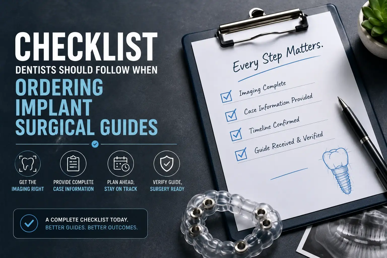

A surgical guide is only as useful as the information it was designed from. Incomplete submissions, poor imaging, missing case details, and rushed timelines are the most common causes of guides that do not meet clinical expectations, and they are all preventable with a structured pre-submission checklist.
This article provides a comprehensive checklist for dentists ordering implant surgical guides, covering every step from imaging acquisition through guide receipt and pre-surgical verification. Follow this checklist on every case and guide-related problems become the exception rather than the norm.
Part 1: Imaging Checklist — Before You Scan
CBCT Imaging Requirements
Confirm the following before acquiring your CBCT:
- Field of view covers the entire implant site plus at least 10mm beyond in every direction
- Full arch FOV of 14x14cm or larger confirmed for full arch or multiple-implant cases
- Scan protocol set to 0.5 to 1.0mm slice thickness, not the standard diagnostic 1.5 to 2.0mm setting
- Patient in natural head position with centric occlusion before acquisition begins
- Patient briefed on the importance of remaining completely still during the 10 to 20 second scan
- Metal artifact reduction protocol activated if the patient has significant existing metalwork
After CBCT Acquisition
- Review the scan on-screen before the patient leaves, do not assume quality
- Verify that the nerve canal is clearly visible and its path is unambiguous
- Verify that the sinus floor (for maxillary cases) is clearly defined
- Verify that adjacent roots are visible without significant artifact
- If quality is inadequate: rescan before dismissing the patient
- Export DICOM files and confirm file integrity (files should open and display correctly)
Intraoral Scan Requirements
- Capture the full arch (second molar to second molar) including the implant site and adjacent teeth
- Capture the opposing arch
- Capture a bite registration (arches in occlusion)
- Include soft tissue detail around the implant site, gingival margins must be visible
- Verify complete coverage before patient dismissal, no holes in critical areas
- Export as STL and confirm the file opens correctly in your scanner software
Part 2: Case Information Checklist — Before You Submit
A complete case information form prevents the most common submission problems. Confirm you have documented:
Patient and Case Identification
- Patient identifier (initials, ID number, or system reference, no full name required for HIPAA compliance)
- Patient date of birth
- Surgery date (or target date range if surgery is not yet scheduled)
Implant Specifications
- Implant system name, be specific (e.g., Nobel Biocare Active, Straumann Bone Level, Zimmer Tapered Screw-Vent)
- Implant diameter preference (if predetermined)
- Implant length preference (if predetermined)
- Drill protocol version, some systems have multiple drill protocols; confirm which applies
Clinical and Surgical Information
- Tooth number(s) for each planned implant
- Number of implants (each implant position must be specified)
- Surgical approach: flapless, limited flap, or open flap
- Any known anatomical concerns (thin buccal plate, nerve proximity, sinus proximity, previous bone graft)
- Anticipated bone quality (if known from previous surgery or other imaging)
Prosthetic Information
- Restoration type: single crown, bridge, implant-supported denture, immediate loading provisional
- Retention type: screw-retained or cement-retained
- Any specific emergence profile or angulation requirements
- Whether immediate loading is planned (if yes, this significantly affects guide design requirements)
Part 3: Submission Timeline Checklist
Timeline failures are the most common reason for rushed or missed surgical guides. Build these dates into your scheduling system when you confirm an implant surgery appointment. Our standard delivery is 5 to 8 business days from submission. View our surgical guide pricing and timeline details.
- Surgery date confirmed and in the calendar
- Guide submission target date set: 3 weeks before surgery minimum
- Imaging appointment scheduled at least 4 weeks before surgery
- CBCT and intraoral scan completed and files exported
- Case information form completed in full
- Files uploaded and submission confirmed received by the lab
- Expedited fabrication arranged if timeline is tighter than 3 weeks (contact lab before submitting)
Do not rely on memory or informal notes for this timeline. Add calendar reminders: one for the imaging appointment, one for the submission deadline, and one to confirm guide receipt before surgery.
Part 4: Guide Receipt and Pre-Surgical Verification Checklist
When the guide arrives, verify the following before storing it for surgery:
Packaging and Sterilization
- Sterile packaging is intact with no tears, moisture, or damage
- Sterilization indicator strips show color change confirming sterilization conditions were met
- Expiration or sterilization date label is present and within the acceptable window (30 days from sterilization)
- Patient identifier on the label matches your case record
Physical Inspection
- No visible cracks, chips, or delamination of the guide body
- All drill sleeves are present, securely seated, and not rotated
- Inspection windows are open and correctly positioned
- Retention features appear complete and undamaged
- Guide appears to match the anatomy size you expected for this patient
Document and File Review
- Any design notes or special instructions included with the guide have been reviewed
- Drill sequence documentation confirms the correct system and drill sizes for your implant
- If design review was requested, confirm the final guide matches the approved design
Part 5: Day-of-Surgery Verification Checklist
On surgery day, before the patient is seated:
- Correct drills for the specified implant system are in the sterile surgical tray
- Drills match the system and protocol specified in the guide documentation
- Saline irrigation is prepared and functional
- Surgical assistant has been briefed on guide seating verification protocol
Before any drilling begins, the guide must be verified as fully seated. This is the single most important intraoperative step for achieving the precision your custom implant surgical guide was designed to deliver.
- Guide seating visual check: no gaps between guide and anatomy from any viewing angle
- Guide seating tactile check: bilateral gentle finger pressure produces no rocking in any direction
- Retention pins inserted and fully seated (if guide design includes pins)
- Second visual check after pin insertion confirms guide remains fully seated
Only begin drilling after confirming all seating check items. A guide that is not fully seated should not be used, pause and investigate the cause before proceeding.
Post-Surgery Documentation Checklist
- Implant system and body type documented in patient record
- Implant dimensions (diameter, length) documented
- Implant placement depth and torque documented
- Guided Excellence noted as guide lab in operative record
- Any seating issues, intraoperative findings, or deviations from plan documented
- Post-operative periapical radiograph taken and archived
Post-surgery feedback is also valuable. If the guide seated perfectly and the case went smoothly, that is useful information. If there were any concerns, sharing them helps us improve designs for future cases. Contact our team through our case submission and support page.