
Surgical guide accuracy depends on a chain of precision decisions starting with the very first clinical step: capturing the patient's anatomy. For decades, this step relied on physical impressions, a process that introduced distortion, delay, and variability before any digital planning began.
Intraoral digital scanners have changed this. By capturing precise three-dimensional surface geometry directly from the patient's teeth and soft tissues, they eliminate the distortion sources inherent to impression materials and deliver a digital dataset that is immediately usable for surgical guide design.
This article explains how intraoral scanning works, why it improves guide accuracy compared to conventional impressions, what scanner characteristics matter for guide applications, and how to get the best results from digital scanning in your guided surgery workflow.
The Problem With Conventional Impressions for Guide Design
Traditional polyvinyl siloxane (PVS) or alginate impressions involve material that flows into the oral cavity, sets, is removed, and then is used to pour a stone model. Each step introduces potential error:
- Material distortion during setting: impression materials shrink or expand as they polymerize
- Removal distortion: pulling the set impression out of undercuts deforms fine details
- Stone model expansion: die stone expands 0.1 to 0.3% during setting
- Model scanning: converting a physical model to digital requires a laboratory scanner with its own accuracy limitations
- Time delay: the impression-to-model-to-scan pipeline takes 24 to 48 hours, during which dimensional stability can vary
Cumulative error from these steps can reach 0.3 to 0.8mm before the guide design process even begins. For a precision device designed to deliver implants within 0.5 to 1.0mm of the plan, this pre-design error is clinically significant.
How Intraoral Scanners Work
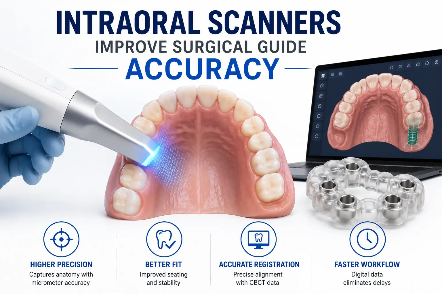
Modern intraoral scanners use structured light projection or confocal laser technology to capture thousands of optical measurements per second across tooth and tissue surfaces. Proprietary algorithms stitch these measurements into a coherent three-dimensional digital mesh, a point cloud that is processed into a watertight STL surface model.
A full-arch scan captures:
- Crown surfaces (occlusal, buccal, lingual) of all remaining teeth
- Interproximal contacts and embrasures
- Gingival margins, papillae, and free gingival contour
- Edentulous ridge surfaces and soft tissue topography
- Opposing arch in occlusion via a bite registration scan
The result is a digital replica of the patient's intraoral anatomy accurate to 10 to 50 micrometers depending on scanner model and technique, a level of precision that physical impressions cannot reliably achieve.
The Accuracy Advantage: Scanner vs. Impression for Guide Design
The accuracy improvement from switching to intraoral scans directly improves the final guide's clinical fit. When Guided Excellence receives intraoral scan STL files alongside CBCT DICOM data, we are working from source data that reflects the patient's actual anatomy without impression or laboratory processing artifacts. Our dental surgical guide design team relies on this precision to deliver seating surfaces that fit passively on first try.
| Factor | Conventional Impression | Intraoral Scanner |
|---|---|---|
| Cumulative Error | 0.3 to 0.8mm | 0.01 to 0.05mm |
| Data Availability | 24 to 48 hours | Immediate |
| Guide Seating Fit | Variable, more rocking | Consistently passive |
| Registration Accuracy | Moderate | High |
| Submission Method | Physical model shipping | Electronic file upload |
The Critical Role of Registration Accuracy
Intraoral scans and CBCT datasets are captured separately with different equipment and different coordinate systems. Before surgical guide design can begin, these two datasets must be aligned, a process called registration or fusion. The accuracy of this registration directly determines how well the guide positions implants relative to the patient's actual bone anatomy.
Registration algorithms identify corresponding surface features, tooth cusps, occlusal anatomy visible in both the CBCT and the intraoral scan, and align them. The quality of this alignment is reported as mean surface distance between matched points. Values below 0.3mm indicate excellent registration; values above 0.5mm indicate misalignment that must be corrected before design proceeds.
High-quality intraoral scans with complete coverage of tooth surfaces provide more registration reference points, enabling more accurate automatic alignment. Partial or low-quality scans with missing areas or mesh noise create registration uncertainty that propagates through the entire design.
Scanning Technique: What Makes a Guide-Quality Scan
Complete Coverage
For surgical guide purposes, complete arch coverage is essential. A full-arch scan should include all teeth from second molar to second molar on both arches, all soft tissue margins in the implant region, the edentulous ridge with soft tissue detail, and a bite registration capturing the arches in occlusion.
Tissue Management During Scanning
The intraoral scanner cannot distinguish between tissue and tooth surfaces, it captures whatever surfaces are in its field of view. Tissue displacement using retractors during scanning improves capture of gingival margins and prevents tissue artifacts on tooth surfaces. Remove excess saliva and blood before scanning as these create optical reflection artifacts.
Implant Site Coverage
The edentulous site itself requires deliberate scanning attention. Multiple overlapping passes from different angles capture the three-dimensional ridge geometry that defines where the guide will seat. Insufficient coverage of this area creates holes in the mesh that compromise guide seating surface design.
Verification Before Submission
Before exporting and submitting scans, verify that the digital model has no obvious holes in critical areas, that gingival margins are captured clearly, and that the file opens correctly in the export format. When submitting to Guided Excellence through our surgical guide case submission process, include notes about any areas of the scan that were challenging to capture.
Scanner Selection: What to Look for in a Guide-Compatible Scanner
Any modern intraoral scanner that exports STL files is compatible with surgical guide design workflows. Specific features that support guide-quality scans include:
- Full-arch accuracy of 25 to 50 micrometers or better with no systematic distortion over the full arch
- Real-time mesh visualization that identifies scan gaps during capture, allowing immediate correction
- STL or OBJ export format compatible with planning software
- Bite registration capability for capturing maxillomandibular relationship
Leading scanners including 3Shape TRIOS, Medit i700, and Ineos X5 all meet these criteria and are routinely used to produce guide-quality digital impressions.
When Scanners Cannot Replace CBCT
Intraoral scanners capture surface anatomy only. They cannot image below the gingival margin, bone anatomy, root positions, the nerve canal, and sinus floor are all invisible to optical scanning. For surgical guide design, intraoral scans must always be combined with CBCT. Neither dataset alone is sufficient.
The combination of high-quality CBCT and high-quality intraoral scan creates the complete three-dimensional picture that surgical guide design requires: bone anatomy from CBCT, surface anatomy from the intraoral scan, merged through registration. This is the foundation of every guide we fabricate at Guided Excellence. See our complete guide services at our implant surgical guide lab.