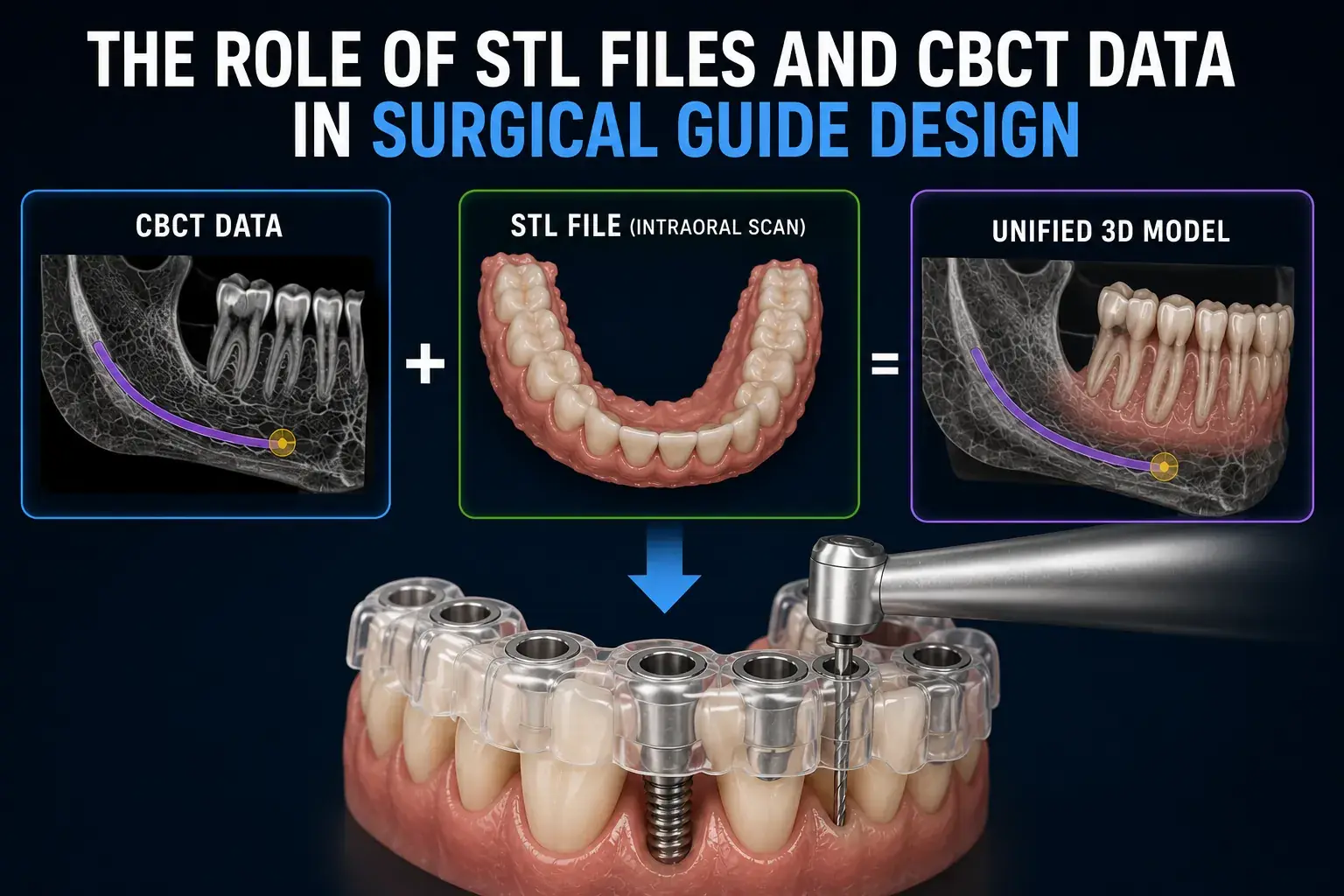

Every surgical guide begins as two separate digital files captured at different times with different equipment: a CBCT scan stored in DICOM format, and an intraoral scan stored in STL format. Neither file alone is sufficient for guide design. Together, when properly aligned through registration, they form the complete three-dimensional patient model that guides the entire design process.
CBCT Data: The Sub-Surface Blueprint
What CBCT Captures
Cone beam computed tomography captures the three-dimensional internal anatomy of the jaw. The CBCT dataset provides bone anatomy in three dimensions including cortical plates and ridge contours, the inferior alveolar nerve canal's complete three-dimensional path, maxillary sinus anatomy and floor height, positions and angulations of adjacent tooth roots, bone density Hounsfield Unit values, and pathological findings such as cysts or bone defects.
DICOM Format
CBCT data is stored in DICOM format, the international standard for medical imaging. A CBCT DICOM dataset consists of hundreds to thousands of individual slice files representing cross-sections through the jaw, typically 0.1 to 0.4mm apart. Planning software stacks these slices and reconstructs the three-dimensional volume for implant planning.
What CBCT Cannot Capture
CBCT does not accurately capture tooth crown surfaces, gingival margins, or soft tissue contours. For surgical guide design, we need both: CBCT provides the bone data, while the intraoral scan provides the surface data that determines guide fit.
STL Files: The Surface Blueprint
What Intraoral Scans Capture
Intraoral digital scanners capture the three-dimensional surface of teeth and oral soft tissues. The STL output provides tooth crown surfaces with precise geometry, interproximal contacts, gingival margins that define emergence profile depth reference, soft tissue topography around the implant site, edentulous ridge surface geometry that the guide must conform to, and the bite relationship between upper and lower arches.
What STL Cannot Capture
Intraoral scans capture surface anatomy only, they cannot image below the gingival margin. Bone depth, root anatomy, nerve canal position, and sinus floor are invisible to optical scanning. This is why CBCT is irreplaceable. Neither file type alone provides a complete picture; only their combination does. Our dental surgical guide design team works exclusively with the combined CBCT and STL model.
Registration: Merging the Two Datasets
The Registration Process
CBCT and intraoral scan data are acquired with different equipment, at different times, and in different coordinate systems. Registration algorithms identify corresponding features visible in both datasets, typically tooth crown surfaces visible in both the CBCT volumetric data and the intraoral scan, and align them using surface matching algorithms. Registration accuracy is quantified as mean surface distance between corresponding points in the two aligned datasets.
Values below 0.3mm indicate excellent registration. Values between 0.3 and 0.5mm are acceptable. Values above 0.5mm indicate registration problems that require manual correction or data reacquisition.
Why Registration Quality Determines Guide Accuracy
A registration error of 0.5mm shifts the entire guide design by 0.5mm relative to the patient's actual bone anatomy. If the virtual implant was planned 2mm from the nerve canal in the registered model, but the registration has a 0.5mm error, the actual distance is 1.5mm, potentially insufficient. This is why registration quality is verified before any design decisions are made.
At Guided Excellence, cases with registration errors above 0.5mm are flagged and either corrected with manual registration refinement or returned to the clinician for improved scanning. This quality gate is non-negotiable. See our manufacturing and quality process.
The Unified Model: Foundation for All Design Decisions
After successful registration, the unified patient model contains both sub-surface bone anatomy from CBCT and precise surface anatomy from the intraoral scan. Every guide design decision is made using this combined model: virtual implant positioning uses bone anatomy and tooth positions simultaneously, safety margin calculations use nerve and sinus positions from CBCT, and the guide seating surface is designed to conform exactly to the tooth and tissue surface geometry from STL.
Practical Implications for File Submission
- Both files are required, a case submitted with only CBCT or only STL cannot be designed to guide precision
- CBCT quality directly affects safety margin accuracy, inadequate slice thickness compromises nerve and sinus clearance calculations
- STL quality directly affects guide fit, holes in the scan mesh or poor soft tissue capture creates guide seating inaccuracies
- Registration accuracy depends on quality in both datasets, high-quality CBCT and high-quality STL enable better registration
Submit both files through our guided case submission portal and our team will review quality and registration accuracy before guide design begins.