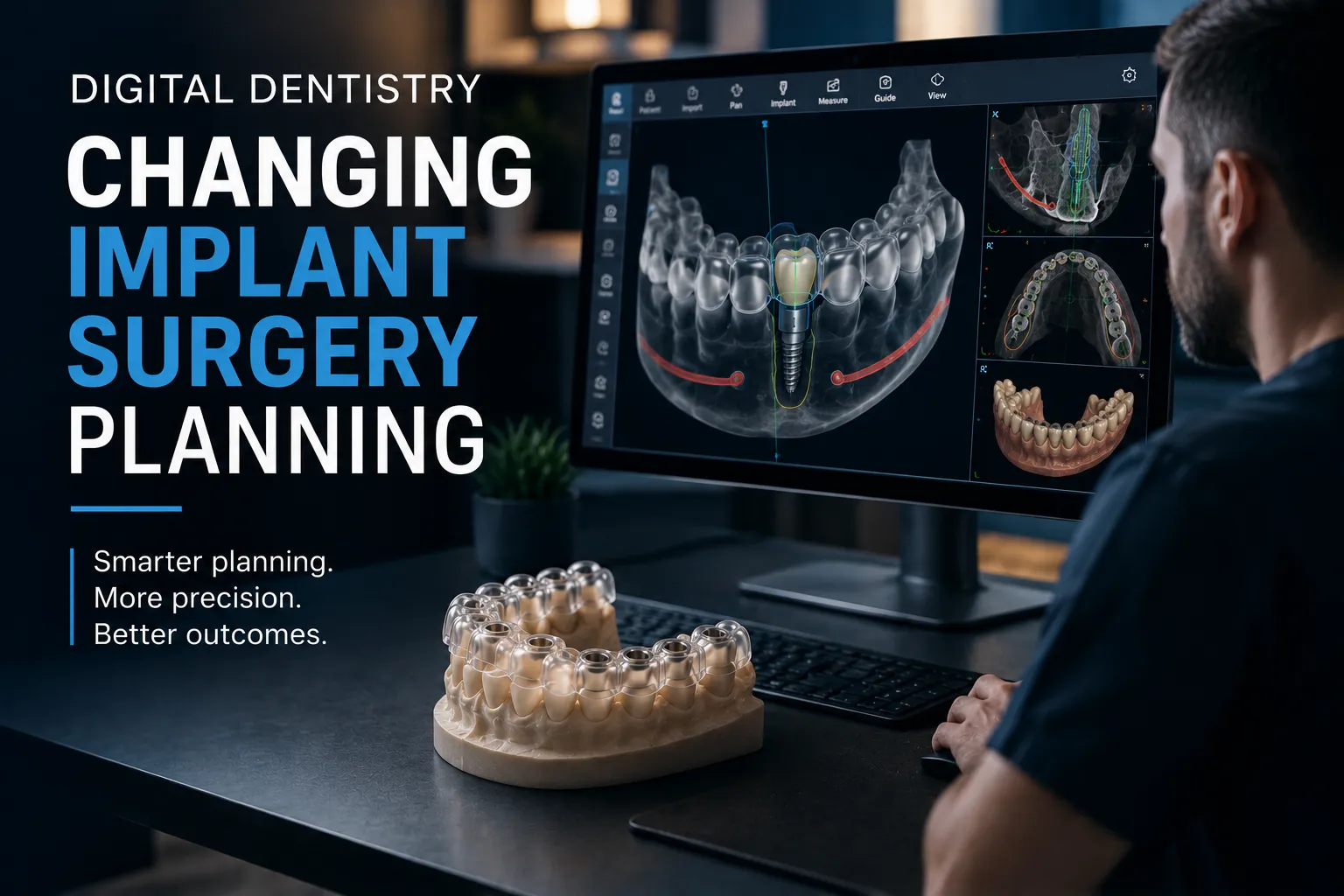

Fifteen years ago, planning a dental implant involved a panoramic radiograph, a set of study models, and a dentist's clinical judgment about where the bone was and where the implant could go. The process was analogue, approximate, and highly dependent on individual skill.
Today, the same planning process can involve a three-dimensional CBCT dataset, a digital intraoral scan, implant planning software that simulates the entire surgical workflow before a drill touches the patient, and a custom surgical guide manufactured to deliver the planned position with sub-millimeter precision.
This is digital dentistry, and its impact on implant surgery planning is not incremental. It is a fundamental change in how implant procedures are conceived, executed, and predicted.
From 2D to 3D: The Foundation of the Digital Shift
The transition from two-dimensional to three-dimensional imaging is the single most significant development in the digitization of implant planning. A panoramic radiograph shows bone height approximately, it represents a flat shadow of a curved, three-dimensional structure. Critical information is obscured: bucco-lingual bone width is invisible, nerve canal depth is estimated rather than measured, adjacent root positions overlap, and bone density is inferred.
Cone beam computed tomography (CBCT) changed this entirely. CBCT captures the jaw in three dimensions with submillimeter voxel resolution. From a single scan, the clinician can measure exact bone height and width at any planned implant site, trace the inferior alveolar nerve canal through its full three-dimensional path, identify root positions of adjacent teeth from any angle, assess bone density, and identify pathology invisible on flat radiographs.
This three-dimensional bone data is the foundation on which digital implant surgical guides are designed. Without it, guide design would rely on estimates rather than measurements, and the precision benefit of guided surgery would be impossible to achieve.
Digital Impressions: Eliminating the Impression-to-Model Gap
In traditional implant planning, the soft tissue and tooth surface data that surgical guides need to seat accurately came from physical impressions poured to stone models, a process with multiple error sources. Impression material distorts during setting and removal. Stone models expand during pouring. Laboratory scanning of physical models introduces additional error.
Intraoral digital scanning replaces this entire chain with a single-step digital capture. The scanner captures tooth surfaces, gingival margins, and soft tissue topography directly as a three-dimensional digital mesh. The resulting STL file is available immediately, in the same digital format that planning software and guide fabrication systems require, with no intermediate analog-to-digital conversion.
The practical effect: guides designed from intraoral scan data fit more accurately and consistently than guides designed from stone models. Registration between the CBCT bone data and the intraoral scan surface data is more accurate when both are high-quality digital captures.
Virtual Planning: Designing the Surgery Before Surgery
Implant planning software is where the transformation in surgical thinking is most visible. These platforms import CBCT and intraoral scan data, register them into a unified three-dimensional patient model, and provide a virtual environment for planning the complete surgical procedure.
In this virtual environment, the clinician can:
- Place virtual implants and see their relationship to bone anatomy, adjacent roots, nerve canals, and sinus floors in three dimensions
- Adjust implant position, angulation, and depth in real time with continuous feedback on safety margins
- Design the final restoration over the planned implant and verify that position supports ideal emergence and crown design
- Simulate the surgical procedure including guide seating, drilling sequence, and depth stops
- Identify potential complications before surgery, nerve proximity, cortical perforation risk, adjacent root concerns, when they can be addressed by plan modification, not surgical revision
This is the prosthetic-driven planning philosophy embedded in our surgical guide design process: the restoration design defines the ideal implant position, and the planning environment verifies that position is achievable safely in the patient's specific anatomy.
The Surgical Guide: From Virtual Plan to Physical Reality
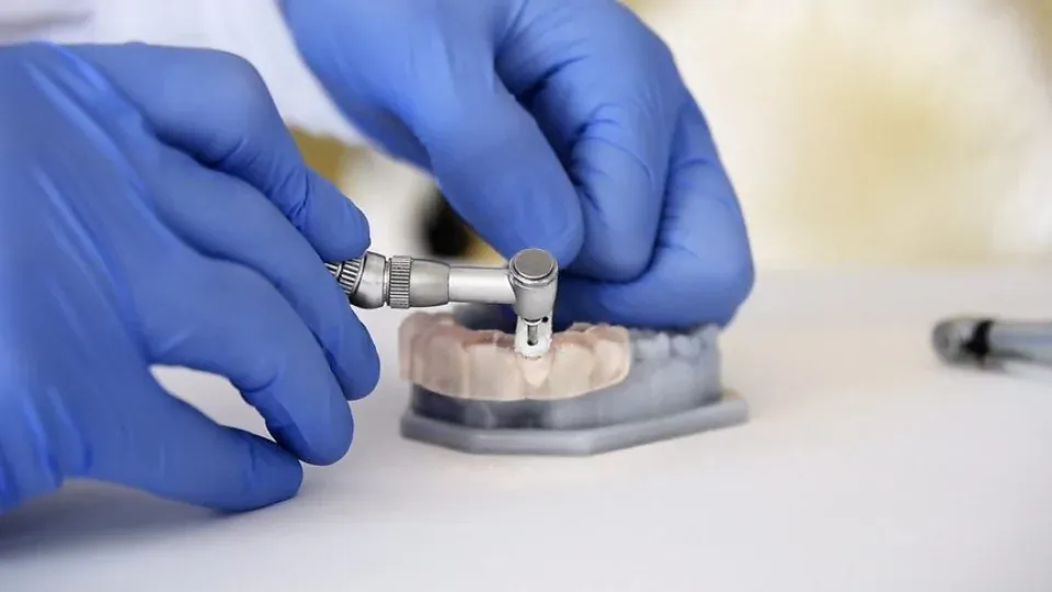
The surgical guide is the device that translates the virtual plan into clinical reality. After the virtual plan is approved, guide design software generates a three-dimensional guide body that conforms exactly to the patient's tooth and tissue anatomy, with precisely oriented drill sleeves that constrain the drill to the approved position, angulation, and depth.
This guide is fabricated using 3D printing technology, stereolithography (SLA) specifically, that produces the designed geometry to plus or minus 0.1 to 0.3mm accuracy. The fabricated guide is verified by seating it on the patient model before shipment, then sterilized and delivered ready for clinical use.
The result is that the plan designed on a computer screen, with full three-dimensional anatomical context, safety margin verification, and prosthetic integration, is delivered to the surgical field in physical form. The surgeon is not executing a freehand approximation of the plan; they are executing the plan itself. That is the clinical transformation that digital dentistry delivers. Explore our implant surgical guide lab capabilities to see how this workflow is implemented.
Digital Communication: Changing How Dentists and Labs Collaborate
Before digital workflows, the dentist-lab relationship in implant cases was mediated by physical impressions, written prescriptions, and phone calls. Information was lost in translation. Assumptions filled gaps. Guides sometimes arrived that did not match clinical intent because the communication chain had broken down somewhere.
Digital workflows make this communication explicit and verifiable. Both parties work from the same three-dimensional patient model. The lab's guide design can be shared as a digital visualization for dentist review before fabrication begins. Specific concerns, implant angulation, guide access for a planned approach, retention pin placement, can be addressed with reference to the actual digital design, not abstract verbal descriptions.
This communication improvement matters most in complex cases where clinical and technical requirements are both high. Full arch restorations, immediate loading cases, and multi-implant esthetic cases benefit most from the shared digital model.
Efficiency Gains: How Digital Transforms Practice Economics
Digital implant planning does not just improve clinical outcomes, it changes the economics of implant practice.
Operative time reduction is the most immediate economic benefit. Guided surgery consistently reduces per-implant surgical time by 15 to 30 minutes. At typical overhead and opportunity cost rates, this translates to $80 to $160 per implant in pure time savings. For a practice placing 50 implants per year, this is $4,000 to $8,000 in annual time savings from the workflow change alone.
Complication cost avoidance is harder to quantify but larger in value. A single nerve paresthesia case, sinus perforation requiring revision, or esthetic failure requiring implant removal can cost $3,000 to $15,000 in direct costs, patient relationship damage, and professional time. Digital planning and guided surgery reduce these events significantly.
Prosthetic efficiency is also enhanced. Guided implants that emerge at planned positions produce first-time prosthetic fit significantly more often than freehand placements. Reduced adjustment time, fewer remake cases, and better patient satisfaction all contribute to practice economics. Our flat-rate guide pricing makes this economic case straightforward, the investment in digital guidance returns multiples in time and complication savings.
Where Digital Dentistry Is Heading for Implant Planning
The current digital implant workflow is already transformative. But active development is moving it further. Artificial intelligence is beginning to automate anatomical segmentation in CBCT data, identifying nerve canals, sinus floors, and tooth roots automatically in seconds. Early AI systems are proposing optimal implant positions based on bone anatomy and prosthetic requirements simultaneously.
Augmented reality systems are beginning to display planned implant positions as real-time visual overlays on the surgical field, potentially adding a feedback layer to guided surgery. These technologies are not yet mature enough to replace static surgical guides, but they are developing toward a future where digital planning is even more tightly integrated with surgical execution.
At Guided Excellence, we monitor these developments continuously and integrate new capabilities when they genuinely improve outcomes. Submit your case to experience what current-generation digital implant planning can achieve for your practice and your patients.